Biopsy Positivity in Mucous Membrane Pemphigoid
Written by Tejah Balantrapu
Published 27th November 2023
In a new study from LVPEI, Drs. Swapna Shanbhag, Sayan Basu, Neha Jain, and Saumya Jakati identify factors that influence conjunctival biopsy positivity in patients with ocular mucous membrane pemphigoid.
Our internal cavities and organs are lined by moist layers of epithelial cells and connective tissue called the mucous membrane, or mucosa. The mucosa forms a barrier, regulating access to the organ. Mucous membrane pemphigoid (MMP) is a rare autoimmune disease in which the body targets its own subepithelial mucosa to produce painful blisters (pustules or pemphigoid) in those membranes, including the eye. Ocular MMP leads to the blistering of the conjunctiva, a mucous membrane that covers the inside of the eyelids and the whites of the eyes. Conjunctival blisters often lead to cicatrization, the scarring and distortion of adjacent tissues. OMMP leads to chronic cicatrizing conjunctivitis (CCC), eventually scarring the cornea and causing blindness. This makes early detection of OMMP crucial for starting immunosuppression therapy and halting the progressive cicatrization of the eye.
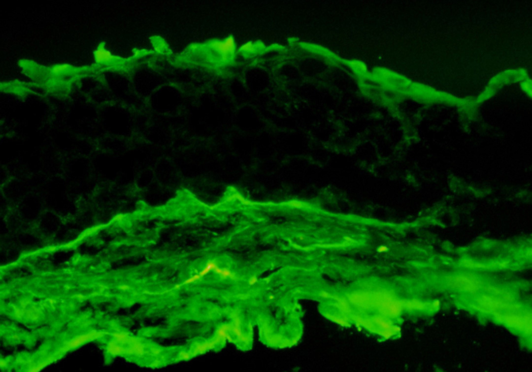
Conjunctival biopsy is the standard test in the MMP diagnosis algorithm. Using direct immunofluorescence (DIF), a tiny portion of the conjunctival tissue is examined for antibodies such as IgG, IgA, IgM, or the proteins C3 and C4—molecular indicators of MMP—that aggregate along the cellular borders. Despite its usefulness, DIF has a wide range of sensitivity (anywhere between 30% and 80%) in conjunctival tissue, making it unreliable for OMMP confirmation. The sample region in the body, the sample size and a host of factors can also impact the test result. Unraveling the factors that impact DIF sensitivity could help improve the accuracy of DIF results.
In a new study published in the journal Cornea, Drs. Swapna S. Shanbhag, Sayan Basu, Neha Jain, and others from LVPEI examined the factors affecting DIF positivity in conjunctival biopsy. This retrospective observational study comprised 61 patients (100 eyes) with OMMP who presented at LVPEI over a four-year period and underwent conjunctival biopsy for DIF in at least one eye. Among them, 54% of patients, or 45% of eyes, obtained a DIF-positive result. A bilateral biopsy (both eyes) was performed on 64% of patients, while 36% of patients underwent a unilateral biopsy (one eye). More than half (59%) of the 39 patients who had a bilateral biopsy came out DIF-positive in at least one eye. On the other hand, of the 22 patients who had one eye biopsied, only 45% tested DIF-positive. Since MMP is a systemic disease, even if one eye reports a false-negative result, a bilateral biopsy can increase the likelihood of a correct diagnosis.
The protein C3 was the most common marker for OMMP observed in this cohort, seen in all DIF-positive eyes, and unlike previous studies that reported the IgG antibody as the most common. The reason for this is unknown, but it could be due to genetic divergence in populations. The study discovered that older patients and those with a severe form of the condition had a lower chance of a positive biopsy. Increased ocular surface scarring can result from both time and disease severity, a probable cause of negative biopsy outcomes. The study emphasizes performing a bilateral conjunctival biopsy in the early stages of the disease to reduce false-negative results.
‘Ocular mucous membrane pemphigoid, lacking proper diagnosis and management, can cause end-stage corneal blindness,’ remarks Dr. Swapna Shanbhag, consultant ophthalmologist at LVPEI and corresponding author of the paper. ‘The diagnosis, although initially clinical, is bolstered by a positive conjunctival biopsy. A false negative result can cost us the window of opportunity where systemic immunosuppression can be used to salvage vision. Hence, it is crucial to get accurate DIF results.’
Citation
Jain N, Jakati S, Shanbhag SS, Basu S. Direct Immunofluorescence Findings and Factors Affecting Conjunctival Biopsy Positivity in Ocular Mucous Membrane Pemphigoid. Cornea. 2023 Oct 3. doi: 10.1097/ICO.0000000000003382.
Photo credit: DIF conjunctival biopsy; by Saumya Jakati; LVPEI.