In a new study Rashmi Deshmukh from LVPEI in collaboration with researchers from Royal Victorian Eye and Ear Hospital and Vision Eye Institute, Australia explore the outcome of collagen crosslinking in patients who have keratoconus along with limbal stem cell deficiency caused by vernal keratoconjunctivitis.
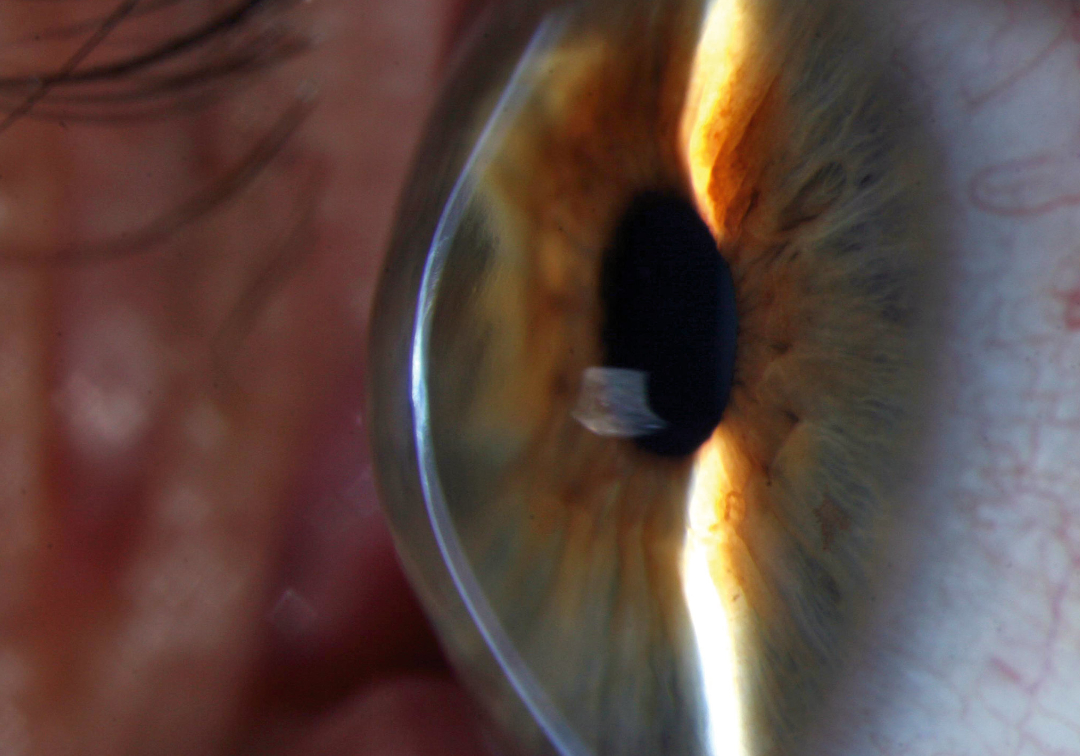
Collagen fibres of the cornea, the eye’s outer layer, give it its transparent, dome shape. When these fibres weaken, the cornea gradually thins and protrudes outward into a cone, affecting vision and causing a condition known as keratoconus (KC). It is a rare but progressive sight-threatening condition; it worsens with time. A major risk factor for keratoconus is vernal keratoconjunctivitis (VKC), an allergic eye disease characterized by itching and eye-rubbing (a well-known contributor to KC). This not only harms corneal structure but also damages limbal stem cells ─ specialized cells present at the corneal rim that help regenerate the outer layer of cornea (the epithelium). This can lead to limbal stem cell deficiency (LSCD), a condition that results in epithelial defects and scarring, which in turn can cause decreased vision and delayed healing of the corneal surface. Together KC, VKC and LSCD create a complex clinical scenario with high risks and challenges to treatment.
Collagen crosslinking (CXL) is the standard procedure used to halt the progression of KC. During CXL the collagen fibres are strengthened using riboflavin (Vitamin B2) which is a photosensitiser that gets activated by ultraviolet A light and sets off new chemical bonds between the collagen fibres. While CXL is effective, performing it in eyes with VKC-induced limbal stem cell deficiency becomes complicated. Standard CXL involves removing the corneal epithelium to allow deeper penetration of riboflavin. In people with deficient limbal stem cells, this step risks delayed healing or persistent epithelial defects (PED). It is therefore necessary to understand CXL treatment response in KC patients presenting with LSCD caused by VKC.
In a new retrospective study published in Clinical Ophthalmology, a team from LVPEI led by Dr Rashmi Deshmukh examined 15 eyes of 11 patients (8 males) that had KC and LSCD who underwent crosslinking. Of the 15 eyes, 14 eyes with partial LSCD underwent epithelium-off procedure, where the corneal epithelial layer is carefully removed before irradiation. One eye with total LSCD underwent an epithelium-on procedure, where this corneal layer is left intact during the process. Preoperative and postoperative data were compared at 1 week, 1 month, 6 months and 1 year.
The study noted that at one year, vision improved in most patients; best corrected visual acuity improved from 0.42 logMAR before treatment to 0.23 logMAR after a year (no vision impairment). The corneal shape also stabilised. The steepest curvature of the cornea (measured as ‘K2’) reduced from 56.2 dioptres before CXL to 54.9 dioptres at one year while the average corneal curvature (Km) decreased from 53.5 dioptres to 52.3 dioptres, indicating a more stable and relatively less cone-shaped cornea. None of the patients required additional limbal cell transplants. The findings indicate that even in complicated cases involving multiple conditions, CXL may be effective.
“Collagen crosslinking can be considered in patients with partial limbal stem cell deficiency and they should be followed up closely for any epithelial healing issues post-CXL,” notes Dr Rashmi Deshmukh, consultant ophthalmologist at LVPEI and the first author of this paper.
Citation
Deshmukh R, Agarwal E, Shanbhag S, Vajpayee RB. Outcomes of Collagen Crosslinking in Patients with Keratoconus and Co-Existent Limbal Stem Cell Deficiency. Clin Ophthalmol. 2025 Sep 12;19:3357-3362. doi: 10.2147/OPTH.S548579. PMID: 40963953; PMCID: PMC12439834.
Photo credit: David Yorston, Community Eye Health Journal, CC BY 2.0