Sohini Mandal, Soumya Sucharita and colleagues from LVPEI report that the severity of Acanthamoeba keratitis is not determined by the amount of organism present but reflects a complex interaction between the pathogen and the body’s inflammatory response.
A contact lens rinsed in tap water, a dive into the swimming pool, or contact with soil are some ways people are exposed to Acanthamoeba, free-living protozoa, found widely in the environment. In some cases, exposure to acanthamoeba can cause serious infections in the brain, skin, sinuses, and eye. In the eye, it invades the cornea (the clear outer layer of eye) causing Acanthamoeba keratitis, a sight-threatening condition that is difficult to diagnose and treat. The infection in its early stages resembles other forms (bacterial/viral) of keratitis resulting in misdiagnosis or a late diagnosis. By the time it is identified, it has already caused significant corneal damage.
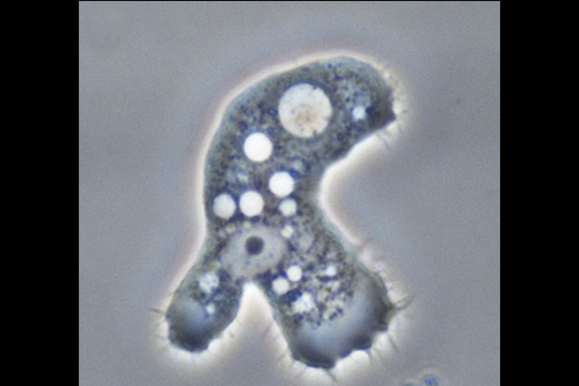
The organism’s biology makes the condition difficult to treat. A key feature enabling this protozoan’s therapeutic resistance is the ability to transition between two forms: the active ‘trophozoite’ form (tropho is Greek for ‘food’—it marks its feeding, growing and invasive phase) that infects tissues; and when the nutrients disappear or drugs are introduced, a dormant ‘cyst’ form. Under unfavorable conditions, trophozoites encase themselves in cysts, double-walled structures that are extremely resistant to external attacks. This capacity for encystment contributes to both treatment failure and disease recurrence. Clinicians often observe that inflammation and tissue damage continue even after the pathogen load (number of pathogens present at the site of infection) has been reduced or the infected tissue has been removed. So, what drives the damage in the cornea - pathogen load or the hosts' immune response?
In a new study published in the Investigative Ophthalmology and Vision Sciences journal, Sohini Mandal and colleagues investigated this question by examining corneal tissue from 23 patients (15 male, 8 female) with Acanthamoeba keratitis over a period of 12 years. The researchers examined the type, location and density of organism present, as well as the severity and pattern of the immune response in the surrounding tissue. A third of the patients had picked up the infection from pond water or vegetative matter. More than half the cases presented with the dormant form reflecting the pathogen’s ability to resist treatment.
In 17 of the 23 cases, the severity of inflammation was independent of the pathogen load, pointing to a complex set of interactions between the two phases of the parasite, and the body’s immune response in damaging the cornea. Younger patients were more likely to have diffuse damage spread across a larger area of the cornea—the causes are unclear. It was noted that at the one-year interval, 65% of corneal grafts had failed, showing the difficulty of restoring vision even after treatment.
“This study highlights that corneal damage in Acanthamoeba keratitis is not simply a function of pathogen burden but is largely driven by the host inflammatory response—underscoring the need for therapeutic strategies that address both infection and immune-mediated injury,” notes Dr Sohini Mandal, the first author of the paper and consultant ophthalmologist at LVPEI Bhubaneshwar.
Citation
Mandal S, Sucharita S, Garg N, Priyadarshini SR, Das S. Host Response and Pathogen Load in Acanthamoeba Keratitis: A Histopathological Analysis. Invest Ophthalmol Vis Sci. 2026 Feb 2;67(2):36. doi: 10.1167/iovs.67.2.36. PMID: 41711507; PMCID: PMC12924137.
Photo credit: Acanthamoeba keratitis, Lorenzo-Morales, Khan, Walochnik, CC BY 4.0