In a new study Katarzyna Rączy, Ramesh Kekunnaya, Brigitte Röder, and other scientists from an Indo-German lab report that the brain requires early visual exposure to develop face recognition. Their data suggests that auditory input might aid the recovery of neural circuits processing visual objects in individuals with reversed congenital cataracts.
The face of a loved one. A hand. A beach, or a cup. As our eyes rest on each of these ‘categories’, specialized parts of the brain process these inputs allowing us to recognize and remember each of them. Information from our eyes travel to a region of the brain called the ventral occipital temporal cortex (VOTC), at the back, and lower part of the brain. The VOTC has a fundamental mapping order. The face-processing sub-regions receive input from the fovea of our retina, the region at the back of our eyes where light-sensitive cells are densest. Our ability to recognize a beach, say, depends on another sub-region, which receives input from the peripheral parts of the retina.
Interestingly, both the face and the place regions of the VOTC can be activated when listening to sound inputs, like laughter, or the sound of water lapping onto a beach. So, people who were born without sight may have tuned their category selective regions of the VOTC with ‘cross-modal’, that is, auditory sensory inputs. Thanks to modern cataract surgery, sight can be restored in some of these people with congenital blindness. What happens then? Will the pathways developed by the non-visual sensory systems interfere with genuine visual processing? Or might the auditory representations aid visual processing?
A new study published in Human Brain Mapping aims to explore whether cross-modal activity aids or interferes with visual input in individuals who had their sight recovered through congenital cataract surgery. The study, by an international team of researchers including Dr. Katarzyna Rączy, Prof. Brigitte Röder, and others from the University of Hamburg and University Maastricht, and Dr. Ramesh Kekunnaya from LVPEI, included eight adult participants (3 males) who were born blind with dense bilateral cataracts but gained sight in infancy (6 to 48 months of age) after surgery (CC group) and eight normally sighted controls who matched for age and sex (SC group). The average age of participants was 42 years. The participants were presented with short (1.8 seconds) video or audio clips of body parts, scenes, faces, and other objects. Then, brain regions that selectively responded to these categories were identified using Functional magnetic resonance imaging (fMRI) scans.
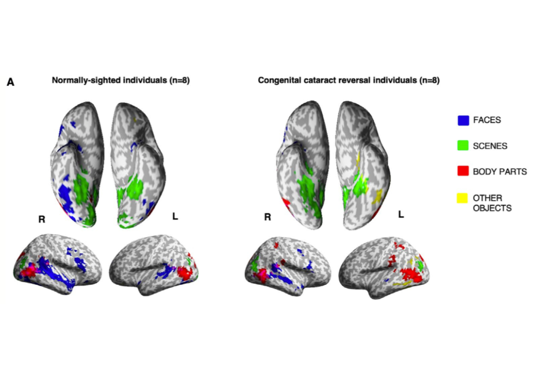
In the CC group, faces elicited markedly lesser selective activation in the relevant brain sub-regions than in the control group. These neural data might explain why people with reversed congenital cataract typically suffer from difficulties in recognizing people by their face. Interestingly, no major group differences were found for the neural circuits processing body parts, objects and scenes. These results suggest that foveal vision is essential to trigger the development of the human face processing system. Interestingly, the authors found evidence that the sound-based representation seemed to have facilitated the activation of the relevant visual category in the VOTC of CC individuals, likely aiding their visual recovery.
'Early foveal vision seems to be crucial for the development of the human face processing system although the fovea itself develops over the first four years. Such 'sleeper' effects highlight the need for early intervention in case of congenital blindness,' notes Prof Brigitte Röder, Head of Biological Psychology and Neuropsychology at the University of Hamburg, and an author on this paper.
Citation:
Rączy K, Linke M, van den Hurk J, Heitmann C, Guerreiro MJS, Zhan M, Kekunnaya R, Goebel R, Röder B. Visual and Auditory Object Representations in Ventral Visual Cortex After Restoring Sight in Humans. Hum Brain Mapp. 2025 Aug 15;46(12):e70316. doi: 10.1002/hbm.70316. PMID: 40851436; PMCID: PMC12375873.
Photo credit: extract from Figure 2; Rączy et al.