Silk film scaffolds
Written by Tejah Balantrapu
Published 27th June 2023
A new paper by Swatilekha Hazra and Dr. Charanya Ramachandran from LVPEI and Souradeep Dey and Dr. Biman Mandal from IIT Guwahati examines the long-term viability of silk fibroin film as a scaffold for culturing corneal endothelial cells. The study determines the impact of silk film’s higher tensile strength and assesses its ability to induce the expression of proteins and extracellular matrix in cultured corneal endothelial cells.
The cornea, a transparent tissue that refracts light into the eye, is made of different layers. The thickest layer is the stroma, lining which are a thin layer of membrane called the Descemet’s membrane and a single layer of cells that hydrates the stroma, called the endothelium. These two layers play a critical role in keeping the cornea clear. Descemet’s is the ‘base’ membrane; it anchors and attaches to the stroma and the endothelium. The endothelium develops at birth as a single layer of cells which are tightly packed. However, the size of the cells expands over a lifetime. This is because dying endothelial cells are not replaced with new cells, rather the old cells increase in size to fill the gap. So, a damaged endothelium needs to be replaced with a transplant; indeed, nearly half of all corneal transplants are conducted to replace a damaged endothelium. However, corneal tissue is scarce, and transplants must factor in disadvantages like immune rejection or graft failure.
One solution to tissue scarcity is engineering a corneal endothelial layer. A variety of synthetic and biological materials can work as a substrate upon which a corneal endothelial cell layer is grown. Films of silk protein (fibroin) are one such substrate material with a lot of promise. Their refractive index is very close to natural tissue. Silk film is non-toxic, biocompatible, degradable at a rate suitable for graft adhesion, and adopts the cornea’s shape, with very high mechanical strength. A previous paper from LVPEI noted all these in vitro attributes of silk film and found two particular varieties of silk that were candidates for corneal endothelial scaffolds. However, silk film’s tensile strength is many times greater than the natural base membrane. This difference may alter cell phenotype (making the corneal endothelial cells behave like fibroblasts thus altering their function). Any such potential change needs careful study, given the dynamic interactions between cells and the network of macromolecules, the extracellular matrix (ECM), they produce to provide biochemical and biomechanical support to each other.
In a new paper in ACS Biomaterials Science Engineering, Swatilekha Hazra and Dr. Charanya Ramachandran from LVPEI and Souradeep Ray and Dr. Biman Mandal from IIT Guwahati examine the long-term cell-matrix interaction and ECM secretion of corneal endothelial cells on a silk film substrate. The study used silk protein sourced from two non-mulberry silkworms, Philosamia ricini (PR) and Antheraea assamensis (AA), to prepare thin (15 microns) films. Corneal endothelial cells were seeded on these films and assessed at 4 time points over 30 days and compared to the cells cultured on fibronectin-collagen 1 (FNC)-coated plastic dishes. The study assessed a specific set of proteins, like laminin and collagen 8 which are unique to the Descemet’s membrane, to proxy ECM health.
Cells cultured on both silk films produced proteins that closely mimicked natural tissue, and in some instances, the films performed better than FNC. In fact, ECM proteins that indicate altered and abnormal behaviour were lesser-the silks’ higher tensile strength and mechanical properties did not negatively impact cell behaviour or morphology. These results point to the suitability of silk film for safely engineering a corneal endothelial layer in the lab.
‘Though this is an oft-overlooked aspect in tissue engineering, it is important to ensure that the material used to culture the cells does not negatively impact their growth, phenotype, or function. This study gives us further confidence that the silk films are indeed suitable for engineering the corneal endothelium for transplantation,’ says Dr Charanya Ramachandran, Scientist, Prof Brien Holden Eye Research Centre, LVPEI.
Citation
In Vitro Profiling of the Extracellular Matrix and Integrins Expressed by Human Corneal Endothelial Cells Cultured on Silk Fibroin-Based Matrices, Swatilekha Hazra, Souradeep Dey, Biman B. Mandal, and Charanya Ramachandran
ACS Biomaterials Science & Engineering 2023 9 (5), 2438-2451
DOI: 10.1021/acsbiomaterials.2c01566
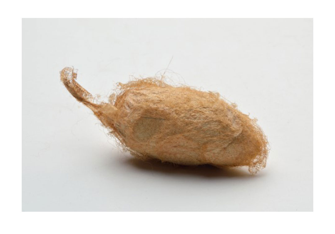
Photo credit: An Antheraea assamensis cocoon; photo by Biswarup Ganguly; CC BY 3.0.