In a new study from LVPEI Deeksha Prasad, Sayan Basu, Vivek Singh, Pragnya Rao and others compare two methods of inducing meibomian gland dysfunction in rabbit models.
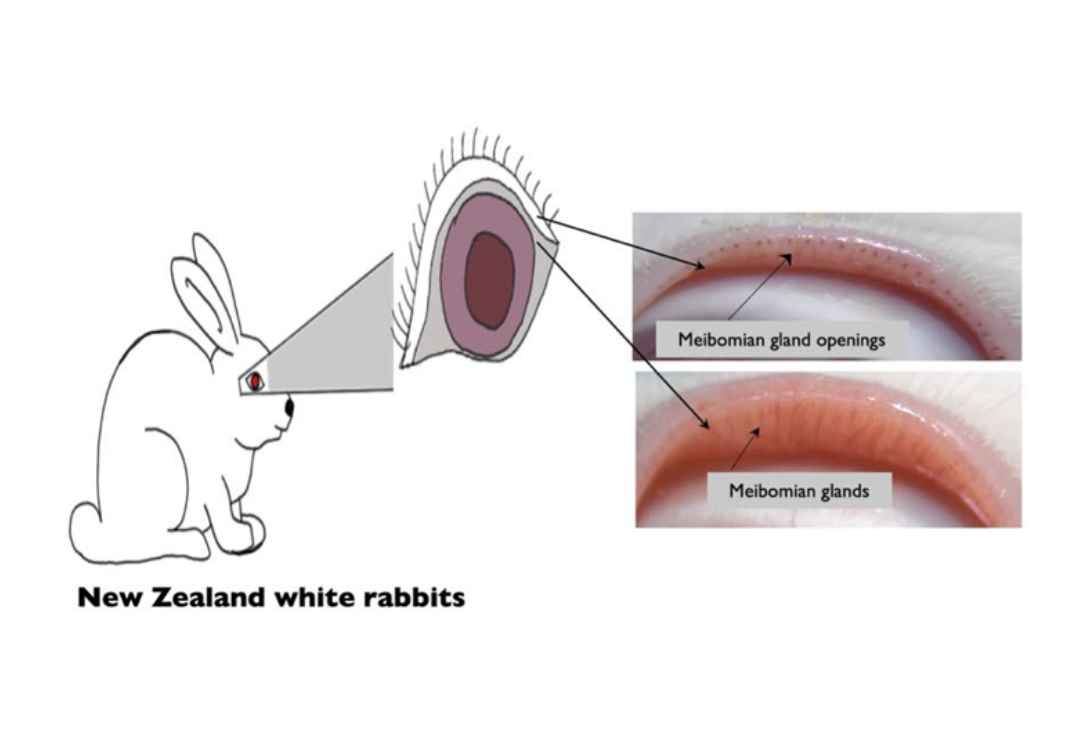
Human tears have 3 parts: an inner layer of mucous, an aqueous (watery) middle layer, and an outer lipid layer called meibum. Our eyelids secrete meibum through oil-secreting tunnels called meibomian glands. Meibum prevents tears from evaporating quickly and maintains the structural integrity of the tear film across the eye. When meibomian glands are blocked or damaged, the lipid layer thins down, the tear film loses stability, and the eye dries out. Meibomian gland dysfunction (MGD) is a leading cause of evaporative dry eye (EDE) disease, in turn the most common type of Dry Eye Disease (DED). Cases of EDE are expected to rise with climate change and digital screen-use, but many risk factors and aspects of meibomian gland dysfunction are unknown.
It is challenging to study MGD in humans and the affected tissue cannot be fully analysed while the patient is alive. This necessitates the need for animal models that can replicate the dysfunction in a research setting. Non-invasive and objective diagnostic assessments such as ‘non-invasive tear break-up time’ (NITBUT), Lipid layer thickness or meibography imaging are common tools to assess dry eyes in humans—but they are seldom used in animal models. Rabbits, with their large eyes and ocular surface, serve as a good model because they allow the use of imaging and diagnostic techniques similar to those used in humans.
In a new paper published in Scientific Reports, a team from LVPEI led by Drs Deeksha Prasad, Vivek Singh, Pragnya Rao and others do just that: assess MGD in a rabbit model. They used two methods to induce MGD in rabbits: chemical cauterization (CC), which blocks gland openings with a chemical agent and electrocauterization (EC), which uses controlled heat to seal the gland openings across the rabbit eyelids. This allowed the study to compare the two major methods of cauterization used in this field. The study included 18 male rabbits (9 in each group) and followed them for 12 weeks, assessing tear film, imaging, tissue and gene expression at days 7, 14, 30, 60, and 90.
The findings showed that tear stability, measured as NITBUT and LLT, decreased significantly in both groups though there was no big change in tear volume (tears are produced by other glands, while meibum maintains stability). The team was able to establish a baseline and dysfunction-induced changes to the meibomian glands in objective terms, which can also help future studies in this field. It was noted that the tissue and gene expression profiles were also similar between the two groups. The study shows that while both methods induce MGD with similar results, CC is more cost-effective and produces gradual changes that resemble the natural course of the disease. EC requires more technical skill, though it produces faster and a more uniform change. These models can help test new diagnostics and therapies that target EDE.
'Chemical cautery of meibomian glands in rabbits is a newer method and more cost-effective than EC,' notes Dr Pragnya Rao, the corresponding author for this study. “This comparison increases the methods available to us for understanding the changes to these glands, and to test newer drugs and diagnostics for dry eye disease.'
'Meibomian gland dysfunction is a key contributor to dry eyes, and our study introduces a simpler rabbit model that goes beyond traditional environmental-based animal models. This new model requires further validation, which is currently underway in our lab using an in vitro cell culture system. The goal is to better mimic the disease biology of dry eye conditions, ultimately improving clinical diagnostics and management strategies,' concludes Dr Vivek Singh, Senior Scientist and a corresponding author of this paper.
Citation:
Prasad D, Jakati S, Bokara KK, Basu S, Singh V, Donthineni PR. Comparative assessment of structural and tear film alterations in rabbit meibomian gland dysfunction models using chemical and electrocauterization techniques. Sci Rep. 2025 Jul 2;15(1):23494. doi: 10.1038/s41598-025-06923-9.
Photo credit: Fig. 1, Prasad et al.