Limbal stem cell deficiency (LSCD) is a condition where the cornea loses its ability to repair itself after trauma. The loss of stem cells in the limbus, a thin, ring-like barrier between the cornea and the conjunctiva, leads to LSCD. Eventually, conjunctival tissue blankets the cornea, making it opaque and leading to vision loss. For improved therapeutic outcomes, it is essential to have a deeper understanding of this condition.
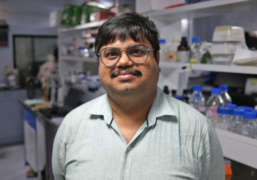
Beyond a pair of biometrically locked doors, in a laboratory at LVPEI’s Kalam Anji Reddy campus, sits Dr. Vivek Singh, principal scientist at the Sudhakar and Sreekanth Ravi Stem Cell Biology Laboratory. Dr. Singh has been studying limbal stem cells, the clinical impact of their depletion, and ways of remediation for over a decade. Along with Dr. Vijay Kumar Singh, a post-doctoral researcher at LVPEI, they discuss their latest research paper on developing a rabbit LSCD model and its role in furthering research in this field.
What got you interested in stem cell research, especially ocular regenerative medicine?
Dr. Vivek Singh: My doctoral research, at the Banaras Hindu University (BHU), was on cancer. The mechanics of cancer stem cells and how they drive cancer progression were hot topics in my field. As the world’s interest in stem cells grew since the early 2000s, so did mine. After completing my PhD in 2009, I moved to the US to begin post-doctoral research at the Cole Eye Institute at the Cleveland Clinic. Over the four years I spent in the US, I developed my expertise on limbal stem cells. Limbal transplantation was already an established procedure at LVPEI and Dr. VS Sangwan was performing many simple limbal epithelial transplantation (SLET) surgeries. But the mechanisms behind SLET’s effectiveness were unclear. When I started working at LVPEI in 2013, Dr. Sangwan and I were involved in a study that established stem cells as the primary factor behind the successful outcomes of SLET. The paper was published in the journal Ophthalmology in 2016. Since then, my lab has focused on stem cell research and its applications in treating ocular surface diseases.
Dr. Vijay Kr. Singh: My doctoral studies were also completed at BHU, where I was involved in elucidation of structure-function aspects of plant defense related proteins using X-ray crystallography and other biophysical tools. After that, I spent more than six years as an assistant professor in Plant Biotechnology at BHU. For more than a year, I was also employed at The Energy Research Institute (TERI). I joined LVPEI in 2019 for my post-doctoral research, to investigate limbal stem cells and their regenerative potential for treatment of corneal wounds.
In this study, you have developed a rabbit LSCD model? Why did you pick a rabbit?
Dr. Vivek Singh: LSCD research employs two animal models: mice and rabbits. A major drawback of mouse models is that their eyes are too small, making it difficult to perform surgery on them. New Zealand White Rabbits have distinctive characteristics such as red eyes and dense white fur due to a lack of melatonin pigment. The lack of pigmentation in these albino rabbits allows for better observation of neovascularization and opacity and makes it easier to study epithelial regeneration post-therapeutic intervention.
Dr. Vijay Kr. Singh: The anatomy of a rabbit's eye is comparable to that of a human, particularly NZ White Rabbits, whose corneas are strikingly identical to human corneas. Furthermore, NZ White Rabbits are easily available and bred at animal facilities for research across India.
Your model uses mechanical debridement to induce LSCD. What is the benefit of using this approach over chemical-injury models that mimic many real-world industrial accidents?
Dr. Vijay Kr Singh: While chemical injury models do mimic real-life chemical burns, the procedure can harm other regions of the eye. Chemical wounds can burn the eyelid or cause an ocular perforation, which can result in further ocular complications. The pathophysiology of LSCD is complicated and includes vascularization, corneal opacity, and conjunctivalization. Studying LSCD is challenging because of the additional problems resulting from adnexal injury (damage to any connected or neighboring tissues). For this reason, we are mechanically injuring just the limbus and depleting the stem cells contained within.
Dr. Vivek Singh: Compared to a chemical damage approach, our model induces LSCD using a more regulated method. Sodium hydroxide is the primary agent employed in chemical injury, and it has a danger of leaking and harming areas of the eye other than the limbus. Our method also has the advantage of being able to generate different grades of LSCD.
Your model can generate different grades of LSCD, but can you control the grade of LSCD induced by this procedure?
Dr. Vivek Singh: We are in the initial stages of exploring this model. Our long-term goal is to assess if we can control the grade of induced LSCD using this procedure. The process has not been optimized yet, but we are optimistic about the possibility.
Dr. Vijay Kr. Singh: Hypothetically, altering the burr size could regulate the degree of LSCD caused by this technique. A 0.5 mm burr, for instance, can be utilized to cause less severe LSCD, but a 2.5 mm burr can cause more severe LSCD. It would be good to pursue such a study in the future.
You note in the paper that a 1.0 mm burr is ‘less traumatic’ when compared to a 2.5 mm burr as was used in a previous study—can you elaborate?
Dr. Vivek Singh: First off, just to be clear, the burr's width and thickness-not its length-are indicated by the values 1.0- and 2.5-mm. Greater trauma will result from a thicker burr, which will destroy tissue across a larger region.
Dr. Vijay Kr. Singh: Because the 1.0 mm burr is more precise than a 2.5 mm burr, there is less risk of ocular damage outside the limbus or to other layers of the eye. Since only the limbal epithelium is being damaged, the procedure is less traumatic-less epithelial tissue is being eroded-versus a thicker burr.
Do you need a qualified surgeon to generate this model?
Dr. Vivek Singh: For optimal precision, we had a surgeon carry out the surgery for this study. However, if the process is established, a non-surgeon too can use it to create LSCD models.
What are some of the benefits and limitations of your model compared to other LSCD models?
Dr. Vijay Kr. Singh: The main benefit of this method is that you can obtain mild, moderate, and severe LSCD animal models without the risk of adnexal damage. The variability in LSCD grade is beneficial for understanding the mechanism underlying the disease and evaluating the limits of regenerative therapies.
Dr. Vivek Singh: Researchers typically begin treatment studies before waiting for the development of persistent LSCD features. It consequently raises the possibility of false therapeutic benefits. So, it is imperative to hold off evaluation of therapeutic intervention for at least four weeks, if not six, for development of persistent LSCD features. Our model's main benefit is that we have serially evaluated LSCD clinical signs and observed a minimum period for LSCD features to stabilize, which may reduce the possibility of a false positive in a therapeutic intervention study.
Can you please elaborate on the value of monitoring gene expression as a tool for understanding LSCD progression?
Dr. Vivek Singh: Gene expression study is useful in understanding the molecular mechanism underlying LSCD progression. Animal models are useful for such studies because there are ethical constraints in using human corneas for similar analysis.
What do you predict will be the impact of your model on future LSCD research?
Dr. Vijay Kr. Singh: Our model is simple to generate, the method is precise, and there is no steep learning curve. Persistent LSCD signs appear in the animal's eye(s) in 4-6 weeks. This means you can start your therapeutic interventional study without having to account for associated complications. I am excited to see how this will change our research output for LSCD models.
Dr. Vivek Singh: Contemporary SLET and Cultivated limbal epithelial transplantation (CLET) surgeries have a success rate between 60 to 80%; about a 70% average. These outcomes could be better if not for delays in medical intervention. We need information on the optimal time to start intervention, so the success rates can be increased to at least 90%. The precise time required for development of persistent LSCD signs with this model can provide an idea on minimum time that may be required for therapeutic intervention. Our ongoing studies with this LSCD model when subjected to allogenic limbal graft transplantation and cultured limbal stem cell transplantation at 4-6 weeks, post a mechanical debridement, have shown an adequate corneal epithelial regeneration. I look forward to all these developments in the near future.
Drs. Vivek Singh and Vijay Kumar Singh spoke to Sayantan Mitra, Science Writer, LVPEI. Read more about their research here.
Citation
Singh VK, Kethiri AR, Pingali T, Sahoo A, Salman M, Koduri MA, et al. Development and validation of a reliable rabbit model of limbal stem cell deficiency by mechanical debridement using an ophthalmic burr. Exp Eye Res. 2023 Sep 26;236:109667.