In a new study, Swatilekha Hazra, Charanya Ramachandran and their collaborators from IIT Guwahati explore the ability of corneal endothelial cells to take topological cues from microscopic shapes on silk fibroin-based substrates for growth and changes in morphology.
The cornea is a multi-layered, outer tissue of the eye that needs to be transparent to let in light. The bulk of the cornea is made up of a layer called the stroma. The innermost layer of the cornea is the corneal endothelium, regulative tissue that hydrates the stroma and keeps it optically clear. This layer expresses a sodium-potassium ‘pump’ enzyme that runs an ion exchange to achieve this critical hydrating function. Binding these two layers is a basement membrane called the Descemet’s membrane. Cells that make up the corneal endothelium are flat, packed together hexagonally (like a honeycomb), and are uniformly sized. The endothelium interacts closely with the basement membrane, responding to cues from the membrane’s 3D shape and structure to alter its own shape or by expressing different types of functional molecules.
Mimicking the topological cues of the basement membrane may prove crucial in engineering an artificial corneal endothelium. A damaged endothelium is one of the main factors that necessitate a corneal transplant. Such artificial tissue then will go a long way in addressing the severe scarcity of transplantable corneas around the world. In previous studies, LVPEI had worked on assembling scaffolds from silk protein (fibroin) to successfully grow human endothelial cells. Would etching patterns and shapes that mimic the basement membrane into these silk film scaffolds improve the growth and morphology of lab-grown corneal endothelial cells?
In a new paper published in the ACS biomaterials Science Engineering journal, Drs Swatilekha Hazra and Charanya Ramachandran from LVPEI and Souradeep Dey and Dr Biman Mandal from IIT Guwahati compare patterned silk film scaffolds to plain ones and report on the differences in endothelial cell morphology and growth. The study introduced two patterns, hexagons and functionally anisotropic (non-uniform) microgrooves, on to scaffolds built from silk sourced from the Antheraea assamensis silk worm. These patterned scaffolds were compared to flat scaffolds of the same silk and tested for a number of physicochemical factors, along with cell adhesion, proliferation, cell density and morphology.
The presence of microgrooves on the scaffolds improved cell adhesion, growth, morphology and maturation when compared to flat or hexagonal patterns. The expression of the sodium-potassium exchange pump protein (NA+K ATPase) was significantly higher in cells supported by microgrooved scaffolds when compared to the other patterns. Similar to previous studies, the patterns did not impact optical transmittance of the scaffolds nor did the tensile strength. The study adds evidence that human corneal endothelial cells respond to topographical cues from their substrates. Further refinement of microgrooves, which are easier to produce than hexagons, should open up better methods of engineering corneal endothelial cells for transplantation.
The field has been keen on developing methods to engineer corneal endothelial cells for transplantation, which will allow us to supplement donor tissue shortage, notes Dr Charanya Ramachandran, Scientist, Prof. Brien Holden Eye Research Centre at LVPEI and the corresponding author of this paper. 'Addition of features that mimic the native substrate is expected to improve the quality of the engineered tissue increasing the post-transplant function and survival. Our study showed that microgrooves significantly increased the quality of cultured cells in terms of their morphology and marker expression, suggesting that their incorporation may be a valuable addition.'
Citation
Hazra S, Dey S, Mandal BB, Ramachandran C. Addition of Microscale Topographies to Silk Fibroin Film Modulates Corneal Endothelial Cell Behavior. ACS Biomater Sci Eng. 2025 Jun 9;11(6):3586-3596. doi: 10.1021/acsbiomaterials.5c00200. Epub 2025 May 14. PMID: 40366209.
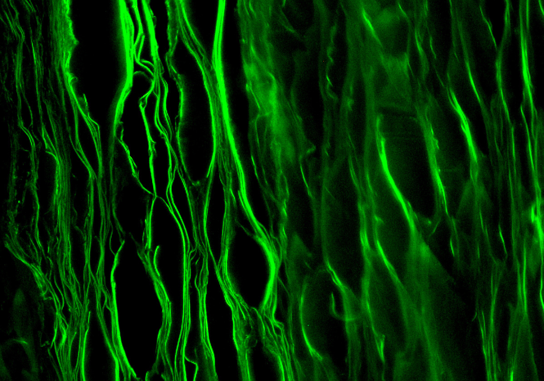
Photo credit: Silk Fibrin lamellar scaffold; Bibhas Kumar Bhunia; CC BY 3.0