People who are born blind can have their remaining senses enhanced by adaptive changes in the brain. The developing brain adapts to the loss of a sense by reorganizing the sensory cortices to improve the processing of the information coming from the intact senses. Among congenitally blind individuals, the visual cortex is known to be activated by auditory input via the auditory cortex and/or the thalamus, the anatomical relay station for most sensory information going to the cortex.
But what happens if a congenitally blind person regains their sight? Would these crossmodal connections withdraw? Or would crossmodal plasticity become an impediment to adaptation?
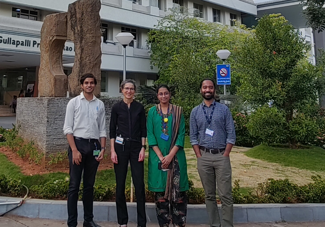
Prof. Dr. Brigitte Röder, the Head of Biological Psychology and Neuropsychology (BPN) at the University of Hamburg, Germany, and Dr. Suddha Sourav, a research engineer at BPN, discuss their latest research paper which sought to answer some of these questions. Their study reports that sound interfered with the earliest visual cortical processing in people who were born blind, but later recovered their sight.
What are your research interests? What led you to have an interest in cognitive neuroscience and crossmodal plasticity?
Dr. Röder: My lab works in the fields of cognitive neuroscience and experimental psychology. We are interested in how the brain develops as a function of experience. To test the role of experience in brain development, we work with the L V Prasad Eye Institute. We investigate people born with severe visual impairment, such as congenital cataracts. Our lab employs electroencephalographic (EEG) recordings, a non-invasive method that uses surface electrodes to record the brain’s electrical activity during perception and cognitive functions.
Numerous cataract surgeries are performed around the world—but their outcomes differ even within the same clinic. Part of this variance arises from different brain states of the patients at the time of surgery. Someone who is born with cataracts will have a different brain organization than someone who develops cataracts later in life and has had visual input shaping visual neural circuits. But how and why? This is where our lab’s basic research found a common interest with LVPEI’s translational research aiming for understanding the neural mechanisms of sight recovery.
Dr. Sourav: My first field of study was electrical engineering. I came to the field of cognitive neuroscience through my second degree in biomedical engineering. Initially I had planned to become a philosopher, but it is tough to carve out a career in philosophy, especially in South Asia (where I am from)! Cognitive neuroscience – analyzing brain activities to understand mental processes – was my closest alternative.
I have two broad interests, one as a scientist and the other as an engineer. As a scientist, I chiefly investigate the electrophysiological activities of the human brain as well as behavioral data to link them to visual/multisensory processing and their development. There are sensitive periods in life, during which experience plays a crucial role in brain development. Loss of experience is much more severe in sensitive periods compared to an equal period of deprivation later in life. I am interested in knowing which aspects of visual processing recover, and to what degree, when vision is restored in individuals who had suffered from a period of congenital blindness.
As an engineer, I work on bringing together and improving upon the existing machines and data processing methods used for this research. To that end, I work as the technical lead at LVPEI’s Indo-German lab. Being an engineer and a scientist puts me in a rare position to work in a very interdisciplinary field alongside doctors, optometrists, and psychologists.
Why is crossmodal plasticity observed in people with congenital sensory loss?
Dr. Röder: The reason for crossmodal plasticity is still not understood. However, we can look at how the brain develops in healthy individuals. Initially, there are excess neural connections, including in the visual cortex, which are later pruned. As the Indo-German lab has documented, this shaping of the visual cortex depends on experience (visual input). The tuning of neural circuits depending on early available environments allows the brain to adjust to the environment it expects to live in. One hypothesis is that crossmodal activation of the visual cortex in congenital blindness originates from some of these excess connections which were not pruned.
Crossmodal activation of visual cortex can be seen in normally sighted individuals too; for example, if we hear a car, we often visualize a car in our minds and such visual imagination is known to activate the early visual cortex. In congenital blindness, the crossmodal activation in visual cortex, however, goes beyond what is observed in healthy individuals.
How did this project begin? What were some of the questions that led to it?
Dr. Sourav: Previous studies have shown that congenitally visually deprived monkeys who regained sight displayed major changes in the extrastriate cortex – visual processing areas of the brain other than the primary visual cortex. For instance, touch activated these visual processing regions of their brain. However, we knew little about how crossmodal plasticity impacts the primary visual cortex in humans. We wanted to know if after vision recovery, the primary visual cortex in humans would process visual stimuli again, and whether the time course of such processing would be typical. In a previous study, we found that after sight recovery, the primary visual cortex seemed to contain a basic representation of the visual field. It processed visual information typically in time, but visual processing in the extrastriate regions was impaired. This study was published in the Journal of Vision.
The next question was what would happen to visual cortical processing in sight-recovered individuals when they were presented audiovisual stimuli instead of just visual stimuli. A functional MRI study by Prof. Dr. Röder and Dr. Maria Guerreiro had found that audiovisual stimuli suppressed the visual areas of the brain. Functional MRI lets you visualize brain activity well in space, but not in time. That raised the question, when does this visual cortex suppression happen? Does it happen in the early stages or is it a general suppression? That is where this study comes in, where we used electrophysiological methods (EEG) for better temporal resolution.
Your study shows that the earliest, stimulus-driven visual cortical activity was suppressed in sight-recovery individuals. How critical is this interference to the functioning of an individual? How does it translate into behavior?
Dr. Röder: The findings of this study are so new that the precise behavioral effects of the earliest visual suppression are yet to be tested. Some researchers have postulated that after sight restitution surgery, rehabilitation should involve both visual and auditory stimuli. It is assumed that the auditory system would guide the visual system in processing visual stimuli. We need to test whether this in fact holds even for individuals who were born blind and recovered sight later in life. We have an excellent opportunity to test this important question for application at LVPEI.
What are the implications of the spatial specificity of visual suppression that you found?
Dr. Sourav: In the group of congenital cataract reversal (CC) individuals, we observed that the earliest visual cortical activity was suppressed more when the sound came from the same side as the visual stimuli. In contrast, in all the other tested groups, irrespective of the presented side, simultaneously presented sounds did not modulate the earliest visual cortical activity. Interestingly, in our simple audiovisual detection task, the CC group’s reaction time did benefit from sounds independent of the presented side. Whether sounds might interfere with the visual performance and interfere more when the auditory and the visual information come from the same side in other tasks is something that we are planning to test.
Can the findings of this study be used to help patients with congenital cataracts who had their sight recovered? If so, how?
Dr. Röder: We know that sight-restored patients do not lose their enhanced auditory abilities. However, the vision of these patients remains impaired if blindness existed at birth. So, their enhanced auditory abilities can help them in their daily life. In the future, we need to compare visual versus audiovisual rehabilitation strategies and see which one is more effective at rehabilitating patients with congenital cataracts who had their sight restored. We are not yet at a stage in the research where we can give recommendations regarding the most effective rehabilitation approaches.
What is the next step for this project? What other projects are you working on?
Dr. Sourav: The next question to investigate is whether the suppression of the earliest visual cortical activity in sight-recovered individuals is limited to the presentation of simultaneous auditory information, or whether other concurrent stimuli (touch, smell, etc.) could also suppress the visual cortex.
We also observed that even with visual cortical suppression there was an overall multisensory improvement in reaction times once sight is recovered. This could be because of sub-cortical brain activity that is aiding in multisensory behavioral improvements. That leads us to the second question: Which tasks are hampered by the suppression of the early visual cortical activity, and which tasks can be done typically? This would tell us the areas that need focus during rehabilitation after congenital cataract surgery.
Dr. Röder: Finding answers to the second question is especially important. From a previous study, we know that lip movement can interfere with the understanding of speech in sight-recovered patients with a history of congenital cataract. Speech sound and the concurrent visual input from the lips are merged in multisensory association areas. Our earlier work had not found typical signs of multisensory fusion in these areas. Whether this is due to visual input arriving late to these areas, or whether the genuine multisensory areas lack multisensory fusion abilities is not known. Understanding the precise brain mechanisms of visual and multisensory processing is not only of high interest for basic science but will be the basis of any evidence-based intervention in vision rehabilitation.
Drs. Brigitte Röder and Suddha Sourav spoke to Sayantan Mitra, Science Writer, LVPEI. Read more about their research here.
Citation
Sourav, S., Kekunnaya, R., Bottari, D., Shareef, I., Pitchaimuthu, K., & Röder, B. (2024). Sound suppresses earliest visual cortical processing after sight recovery in congenitally blind humans. Communications biology, 7(1), 118. https://doi.org/10.1038/s42003-023-05749-3