A Chemical Pack
Written by Soujanya Padikkal
Published 27th August 2026 / Last updated 28th August 2026
A new study from the LV Prasad Eye Institute shows that conjunctival biopsy samples can be transported without immediate freezing, making direct immunofluorescence testing for some ocular surface conditions more accessible in resource-limited settings.
When doctors suspect a case of ocular mucous membrane pemphigoid (oMMP), a biopsy of the conjunctiva is taken. A piece of tissue few millimeters long is removed from the conjunctiva – the transparent layer covering the white part of the eye – and sent to a laboratory.
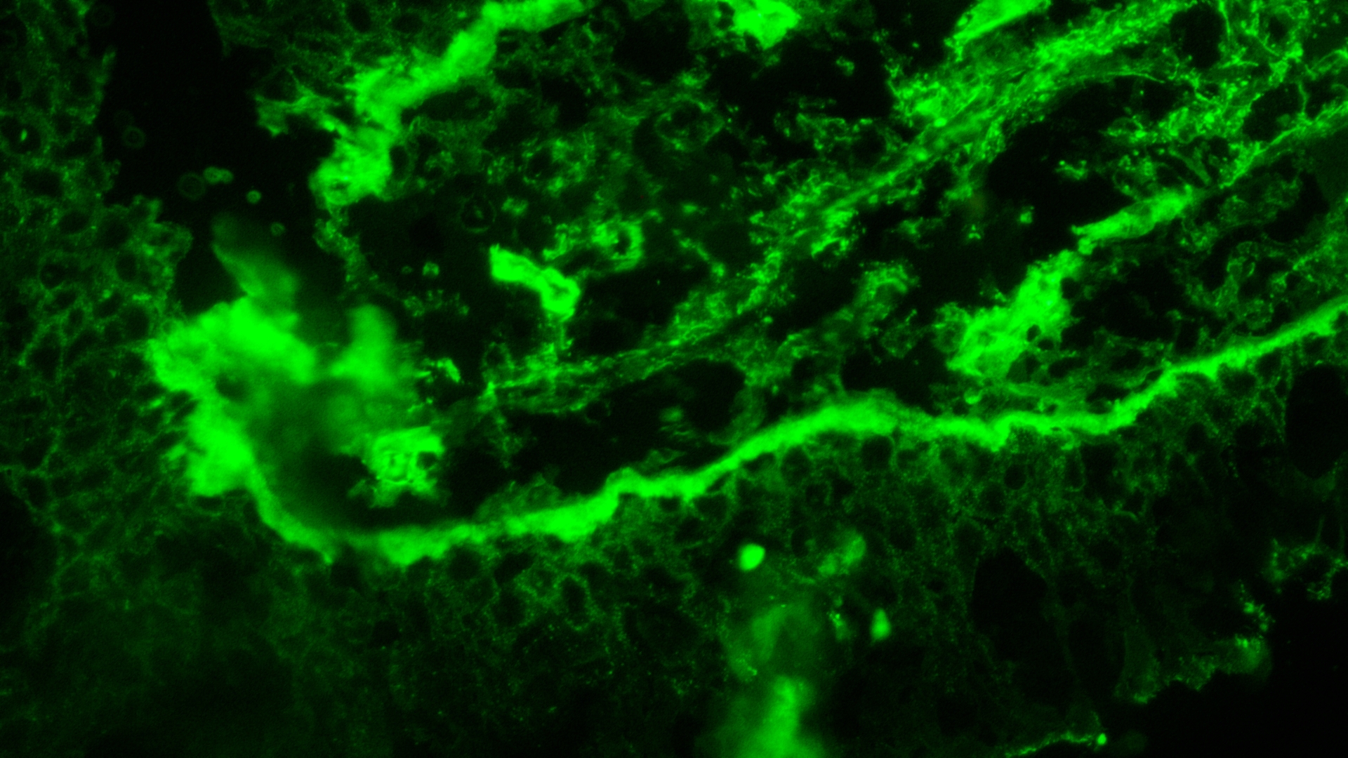
Ocular mucous membrane pemphigoid is an autoimmune disease that causes chronic inflammation, scarring, and can lead to vision loss. It is diagnosed through a test called direct immunofluorescence (DIF), which tests for the unique set of antibodies and immune proteins that target against the basement membrane proteins such as BP180, integrin and laminin that are deposited in the basement membrane of the conjunctival tissues. But for the test to work, the biopsy needs to be snap frozen in liquid nitrogen so that these delicate proteins remain intact and can be detected on the test.
The conventional method is to place the biopsy in an optimal cutting temperature (OCT) compound media, and snap-freeze it with liquid nitrogen immediately. A cryostat microtome; a piece of specialized equipment, is used to cut frozen tissue sections that many eye centres do not have. Patients need to travel long distances to a well-equipped facility, or the samples need to be rushed to one under tight time pressure.
But for the test to work, the biopsy needs to be snap frozen in liquid nitrogen so that these delicate proteins are intact and can be detected on the test
Sometimes this may take days to process; some patients don’t come back for the diagnosis report, and clinicians avoid sending samples due to the limitation of distance and transport. Clinicians are therefore turning to a simpler alternative: using the same OCT media, without immediate freeze drying the sample, and transporting it to a different facility to be processed later. But will DIF still be able to detect proteins in such a scenario?
In a new study published in Cornea, Mariya B. Doctor, Poornima Golthi, Sayan Basu, Saumya Jakati and Anahita Kate from LVPEI investigated whether OCT compounds could be used as transport media for conjunctival biopsies without snap-freezing them. The team analysed 139 biopsy samples from 71 patients suspected of having oMMP.
After collection, each biopsy was placed in a tube filled with OCT, placed within ice packs, and transported 291km to the pathology laboratory, where it was then frozen with liquid nitrogen and processed for DIF. The researchers tracked how long each sample took to arrive, whether the tissue’s basement membrane remained structurally intact, and whether DIF could still detect the immune markers it depends on: IgA, IgG, IgM, and the complement components C3 and C4.
The findings suggest that centers without on-site freezing facilities may not need to process these biopsies locally at all — they could preserve the sample in OCT media, use ice packs, and send it for testing without compromising the result.
The samples preserved in OCT compound and transported in cold chain did not affect DIF results despite an average delay of 4 days. It was noted that 99% of the samples had an intact conjunctival basement membrane, and 43 of the 71 patients showed a positive DIF result. The samples processed within a week and those processed later showed similar DIF positivity rates of 59% and 56% respectively.
The findings suggest that centers without on-site freezing facilities may not need to process these biopsies locally at all — they could preserve the sample in OCT media, use ice packs, and send it for testing without compromising the result. This would be useful for clinicians to aid in the diagnosis of this condition, even in resource-limited settings.
‘A delayed journey for biopsy does not mean a compromised diagnosis. This approach could help overcome one of the logistical barriers to diagnosing oMMP, in resource-limited settings’, says Dr. Mariya B Doctor, Consultant Ophthalmologist, LVPEI and first author of the study.
Citation
Doctor, M. B., Golthi, P., Basu, S., Jakati, S., & Kate, A. (2026). Delayed Processing of Conjunctival Biopsies for Direct Immunofluorescence in Mucous Membrane Pemphigoid in Resource Limited Settings Cornea https://doi.org/10.1097/ICO.0000000000004207
Photo credit: DIF conjunctival biopsy, Dr. Kalyan Rao, LVPEI.