A Pink Glow
Written by Soujanya Padikkal
Published 29th October 2026
Aravind Roy from LV Prasad Eye Institute and researchers from the University of Colorado report that the loss of Rose Bengal fluorescence after green-light activation in bacterial keratitis results from an interplay between the bacteria and corneal-tissue related factors.
In 1882, a Swiss chemist developed a rich, rosy-pink dye to color wool, called ‘Rose Bengal’ (RB), which ended up having interesting applications in ophthalmology. Rose Bengal belongs to a class of dyes that can be ‘excited’ by light of a specific wavelength. In its excited state, RB yields a very reactive type of Oxygen, called Singlet-Oxygen, which is cytotoxic and causes severe oxidative stress that destroys bacterial cells. This property has made researchers explore RB as a potential alternative to treat infections in a world of growing antimicrobial resistance.
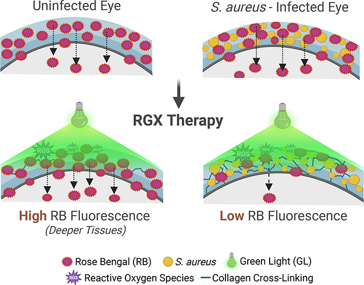
Rose Bengal green-light cross-linking (RGX), a technique developed in 2013, uses green light at a wavelength of 530 nm to activate Rose Bengal, which then produces reactive oxygen species (ROS) like singlet-oxygen. These molecules not only kill microbes but also trigger the formation of new bonds between the cornea’s collagen fibers (the structural proteins that help the cornea maintain its shape and strength).
The activated dye also emits light, a phenomenon known as fluorescence, which can be measured to track the dye in tissues. When the dye interacts with surrounding molecules, its fluorescence reduces. Measuring these changes can provide insight on how effective RB activation is in the infected tissue and whether crosslinking of collagen affects the delivery of antibiotics when used along with it.
Rose Bengal green-light cross-linking (RGX), a technique developed in 2013, uses green light at a wavelength of 530 nm to activate Rose Bengal, which then produces reactive oxygen species (ROS) like singlet-oxygen.
In a new study published in ACS Applied Biomaterials, researchers from the University of Colorado and LVPEI explore this question using an experimental model of S. aureus-infected bovine eyes to study RB’s behavior during RGX. They measured the dye’s fluorescence in different corneal layers and the fluid inside the eye.
They then assessed if the treatment affected corneal strength and the delivery of vancomycin, an antibiotic used for corneal infections. They also conducted in vitro experiments to examine the effect of green light on RB fluorescence and collagen rigidity among other effects.
In vitro experiments showed that this reduction occurred only when bacteria, corneal tissue extract, and light were present together, suggesting interactions between these factors contribute to fluorescence reduction.
The team found that green-light exposure reduced Rose Bengal’s fluorescence by 75-80% in infected eyes, but this was not the case in uninfected eyes. They suggest that RB may bind to the proteins in the infected corneal tissue or form products that do not fluoresce, causing a loss of fluorescence.
In vitro experiments showed that this reduction occurred only when bacteria, corneal tissue extract, and light were present together, suggesting interactions between these factors contribute to fluorescence reduction. Adding ROS scavengers in infected samples containing bovine corneal cell extract prevented this loss, implying that ROS is also involved in fluorescence loss. The treatment, however, did not affect corneal strength or vancomycin delivery.
‘Bacterial infection causes loss of Rose Bengal fluorescence in infected corneas without affecting antibiotic delivery and corneal rigidity’, says Dr. Aravind Roy, Consultant Ophthalmologist, LVPEI. ‘These findings help further explore Rose Bengal’s therapeutic potential in combination with antibiotics for treating bacterial keratitis’.
Citation
George SE, Roy A, Sharma PK, Upadhyay S, Kompella UB. Effect of Green Light-Mediated Corneal Cross-Linking on the Delivery of Rose Bengal and Vancomycin in Staphylococcus aureus-Infected Eyes. ACS Appl Bio Mater. 2026 Aug 22:10.1021/acsabm.6c00339. doi: 10.1021/acsabm.6c00339. Epub ahead of print. PMID: 42638438; PMCID: PMC13592553.
Photo credit: Excerpted from Abstract, George SE et al.