Keratoconus and Contact Lenses: Breaking the Stigma for Better Vision – Kode Venkatadri Chowdary campus, Vijayawada
Published 9th October 2026
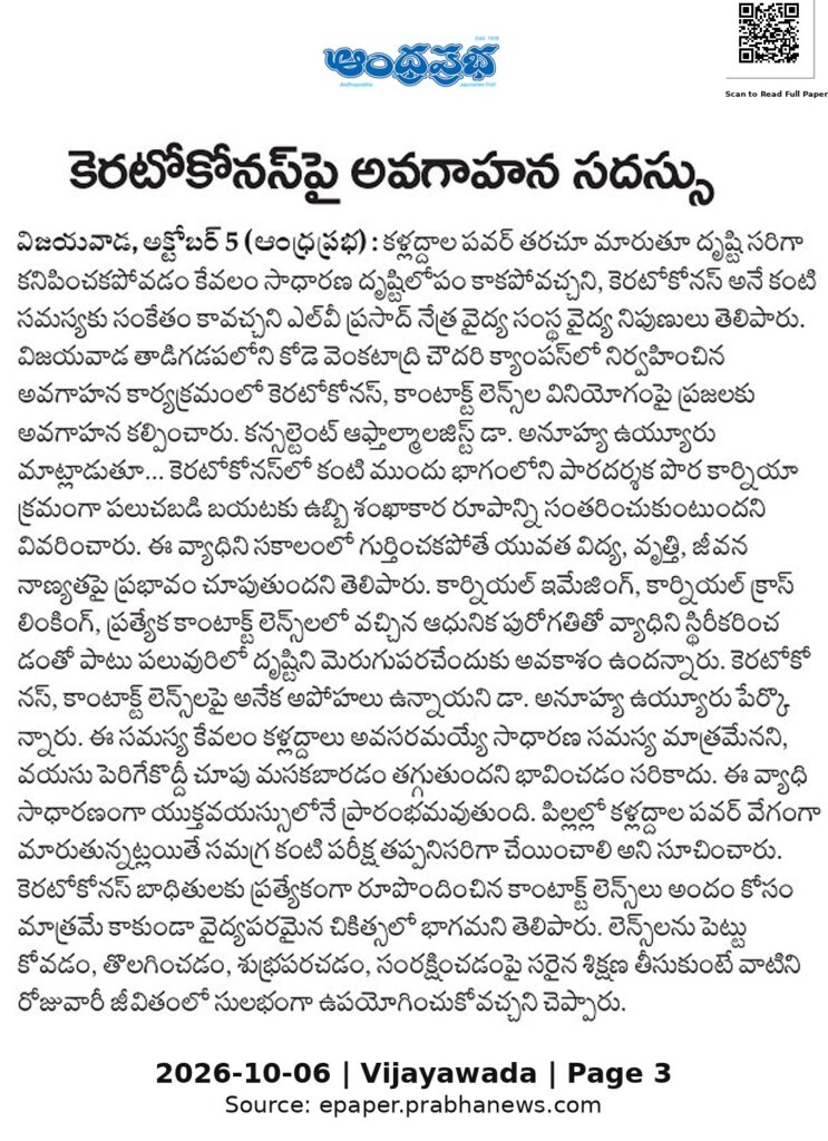
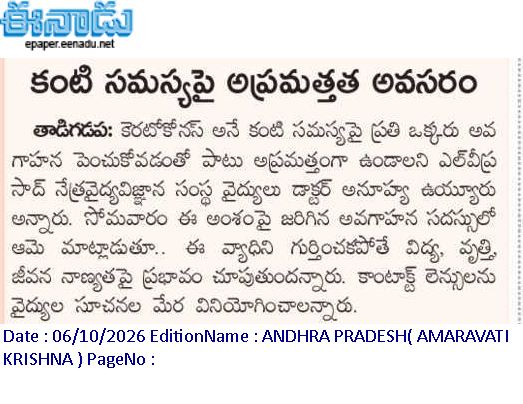
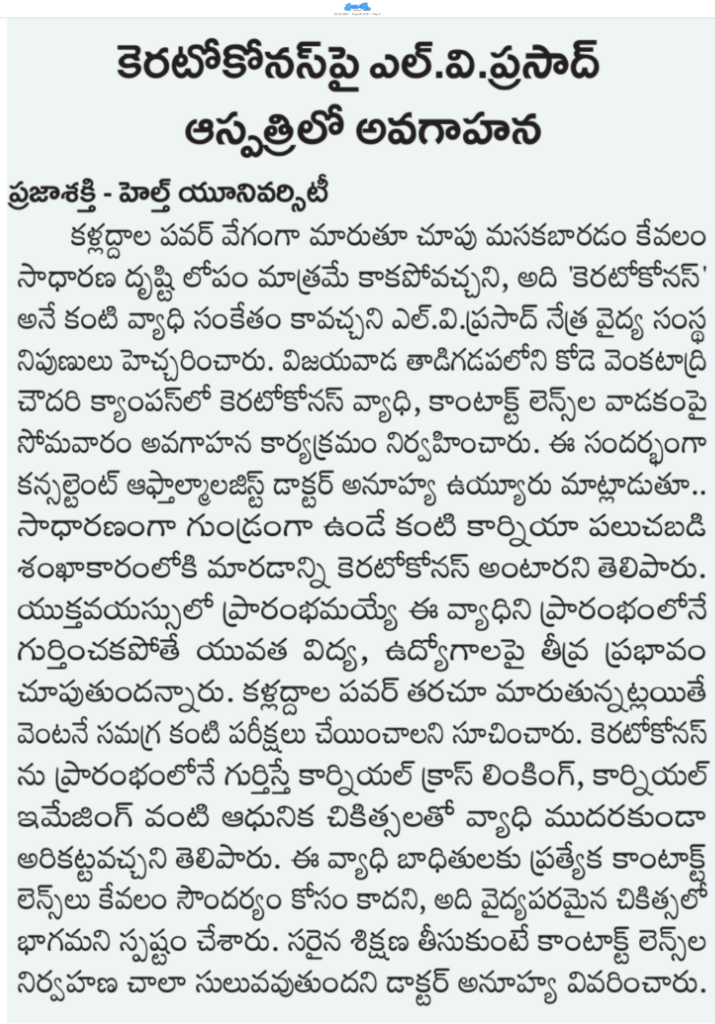
Every year, many teenagers and young adults struggle with frequent changes in prescription glasses and deteriorating vision, often misdiagnosed as simple refractive error or myopia. This may also be the silent onset of keratoconus, a progressive eye condition where the normally round cornea thins and bulges outward into an irregular cone shape. Unlike regular refractive errors, if left undetected, keratoconus can severely disrupt a young person’s education, career, and quality of life. . Delayed care continues to hinder effective treatment.
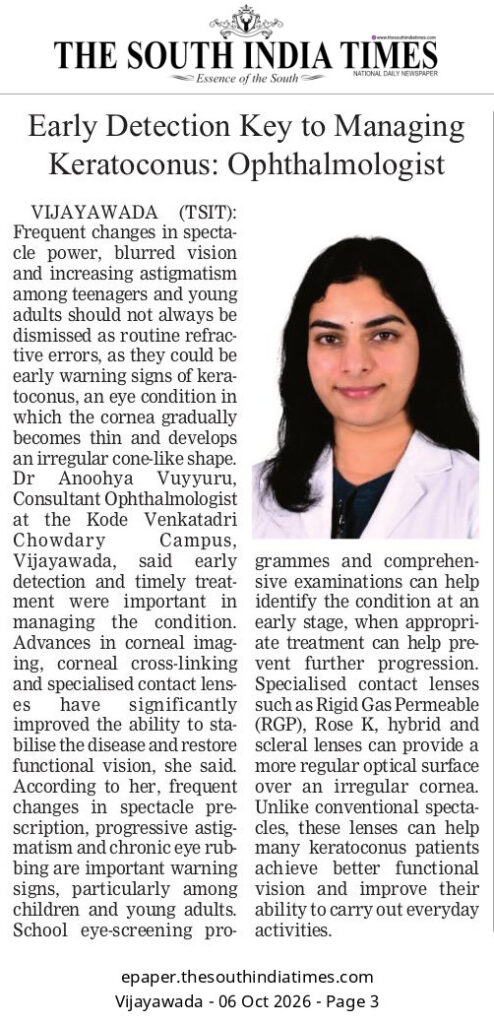
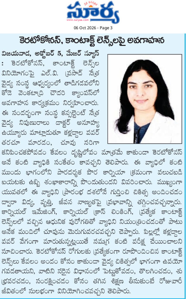
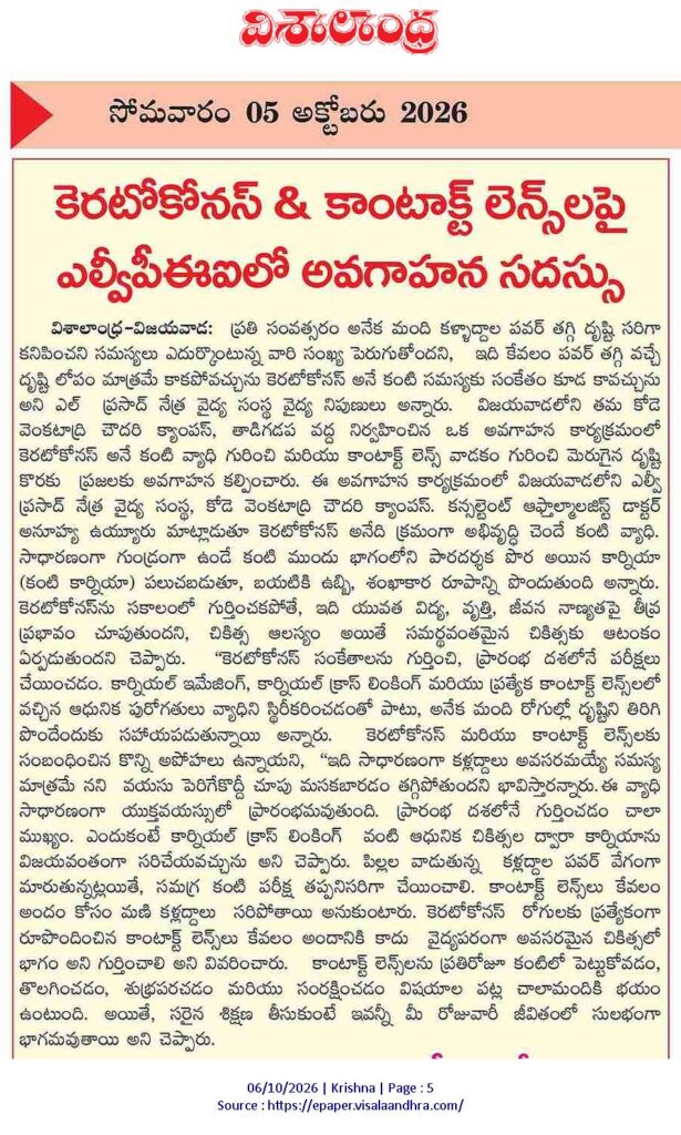
“The key to effective treatment of keratoconus is to recognize the warning signs and investigate them early. Advances in corneal imaging, cross-linking and specialized contact lenses, help stabilize the disease and restore functional vision for many patients.” Says Dr Anoohya Vuyyuru, consultant ophthalmologist at Kode Venkatadri Chowdary campus, Vijayawada.
Early detection of Keratoconus and management with specialized contact lenses can support a near normal lifestyle. Here are few common myths about keratoconus and contact lenses, and the facts every patient and family needs to know.
Myth 1: “It’s just a normal need for glasses; they will outgrow the blurry vision.”
Frequent changes in glasses prescription, progressive astigmatism, or a habit of chronic eye rubbing in children and young adults are major red flags for keratoconus. The condition typically begins in adolescence. Early detection is crucial because modern treatment, such as corneal cross-linking, can successfully stabilize the cornea. School screening can help identify at-risk children before the condition causes irreversible vision loss. If a child’s glasses power is rapidly changing, a comprehensive eye examination is essential.
Myth 2: “Contact lenses are only for cosmetic use; glasses are always enough.”
For keratoconus patients, specialized contact lenses are are a medical necessity. As the cornea becomes increasingly irregular, standard glasses cannot properly compensate for the distorted light entering the eye. Specialized lenses such as the Rigid Gas Permeable (RGP), Rose K, hybrid, or scleral lenses smoothly fit the irregular cornea to create a new, uniform optical surface. For many patients, these lenses restore functional vision and offer major improvement in the activities they can perform independently and the quality of life.
Myth 3: “I won’t be able to handle or take care of specialty lenses by myself.”
Many fear the daily routine of inserting, removing, and maintaining contact lenses. However, proper training makes this a seamless part of your daily routine. At specialized contact lens clinics, eye care professionals provide hands-on training tailored to each patient. They guide you on hygiene, handling techniques, and correct usage. With professional support, patients quickly build confidence, and realize that the dramatic improvement in the quality of vision and life, far outweighs the initial learning curve.
Myth 4: “My vision feels stable right now, so I don’t need regular follow-ups or advanced eye scans.”
While your vision may seem stable day-to-day, keratoconus is a highly unpredictable condition that can silently progress, thinning and steepening the cornea before you experience any noticeable blurriness. Relying solely on how you feel or waiting until you struggle to read a standard eye chart may be dangerous. Structural damage often outpaces your awareness. By the time your vision significantly drops, the disease has already advanced.
Routine, scheduled follow-ups utilizing corneal topography are non-negotiable for anyone diagnosed with or at risk of keratoconus. Topography is a quick scan that maps the microscopic curves and thickness of the front surface of your eye. It acts as a highly sensitive early warning system, detecting minute, sub-clinical changes in the cornea’s shape, long before they impact your daily life. Catching this early progression is the most critical step in keratoconus management because it allows specialists to perform Corneal Cross-Linking (CXL) promptly. CXL is a safe, minimally invasive procedure that uses ultraviolet light and vitamin drops to strengthen the collagen bonds in the eye, effectively halting the progression of the disease. Consistent topographic monitoring ensures specialists can intervene with cross-linking at the right moment, stopping the condition in its tracks, preserving your current visual potential, and preventing the need for complex corneal surgeries in the future.
Every year, we see young patients who have given up on their academic or career aspirations simply because they believed nothing more could be done. Keratoconus does not have to mean a lifetime of poor sight. Do not let stigma or fear define your eye health. Get a comprehensive eye examination done today to avoid complications associated with keratoconus. About LVPEI: Established in 1987, with the vision, to create excellent and equitable eye care systems that reach all those in need,’ the L V Prasad Eye Institute (LVPEI), a comprehensive eye health facility, is a World Health Organization Collaborating Centre for the Prevention of Blindness.
In pursuance of this vision, LVPEI clinician scientists work at the cutting edge of eye research. Through its five-tier ‘Eye Health Pyramid’ and cross-subsidy models, LVPEI provides more than 50% of its services entirely at no cost to the patient, irrespective of the complexity of care needed. LVPEI is among the world’s top eye researchers with over 5,000 published scientific papers in prominent international journals.
Contact: Aneetha Kanukolanu,
Associate Director- Network Communications
Phone: +91 40 6810 2272
Email: aneetha.kanukolanu@lvpei.org
Website: www.lvpei.org