The Eye & Brain Lab
The eye and brain lab at LVPEI investigates how vision and its interactions with the other senses (e.g., hearing or touch) depend on visual experience in early life. This is important because “seeing” may start at the eyes, but vision is also fundamentally linked to the brain. In fact, about one-third of our brain is involved in vision. The proper development of visual neural circuits crucially depends on what a child sees, and how well they can see.
Sight-recovery individuals are a major focus of our lab. Such individuals were born blind, for example, due to dense congenital cataracts, but recovered sight after surgery. Investigating sight-recovery individuals allows us to investigate which visual functions recover after a period of blindness since birth, and to what degree.
Lab Methods: Neuroplasticity, Sensitive Periods, and Vision
The eye and brain lab uses behavioral, electrophysiological, and neuroimaging methods.
Eye Tracking
Eye movements are the fastest human behavior known to us. With eye tracking, these fast, intricate movements can be recorded and analyzed. Using eye tracking, our lab has previously reported that “visual exploration patterns were preserved in individuals with reversed congenital cataract (Ossandón et al., 2022), despite severe residual visual impairments and gaze instability (nystagmus).”

Above: A 900-year-old optical illusion. [1]
Electroencephalography (EEG)
Neurotransmitter release leads to changes of the electromagnetic field in the brain, which appear as weak voltage fluctuations on the scalp. With electroencephalography (EEG), we can record these minuscule voltages, which are about 1,000,000 times smaller than the voltage of a pencil battery! EEG provides information about brain processes with a very high temporal resolution.
Above: Examples of EEG Scans at the lab. [2]
Magnetic Resonance Imaging (MRI)
Magnetic resonance imaging (MRI) detects faint “echoes” of radio signals sent to the brain inside a powerful magnetic field. Using MRI methods we can precisely measure the brain’s structures down to sub-millimeter scale, estimate brain activity via blood oxygenation level changes (functional MRI), and uncover nerve fibers’ structures by following the path of water diffusion in the brain. MRI provides information about brain structures and processes with a very high spatial resolution.
Above: Examples of MRI research at the Eye and Brain Lab. [3]
[1] Stone carving of an ox and an elephant with a common head, at Airavatesvara Temple. Eye movement data overlaid on the image. The red dot is the eye’s current position. The white points are fixationperiods, when the eyes rest on the image. They are punctuated by very fast eye movements called saccades. Video courtesy of Dr. José Ossandón.
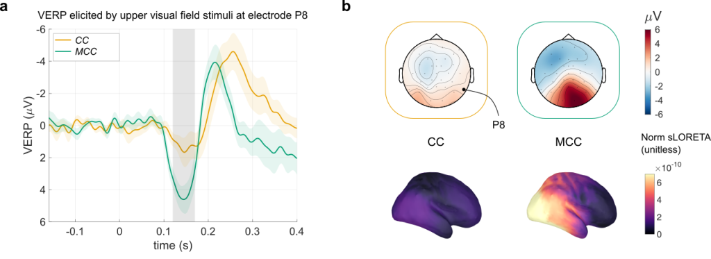
[2] Examples of a. average time-course of the voltage at a posterior electrode after the presentation of a visual stimulus, and b. visualization of the scalp voltages (averaged between 125 – 175 ms) as topography (top panels) or brain source maps (bottom panels). CC: congenital cataract reversal individuals, MCC: matched control individuals for the CC group. Figure adapted from Sourav et al. (2026)
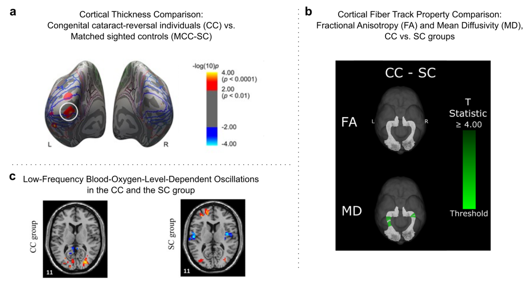
[3] a. Congenital cataract reversal individuals (CC) have been reported to exhibit a higher cortical thickness, Hölig et al. (2022). b. A recent article from Hassett et al. (2026) found specific changes as well as substantial recovery of white matter microstructure in CC individuals. c. Rączy et al. (2022)have reported changes in spontaneous brain activity in CC individuals using functional MRI methods.
Principal investigators
Ramesh Kekunnaya
Dr Ramesh Kekunnaya is the Director of both the Child Sight Institute and the Center for Technology Innovation, and the Head of the Jasti V Ramanamma Children’s Eye Care Centre at LVPEI.
His core clinical interests include simple and complex strabismus in adults and children (squint), childhood cataract, vision development, myopia, amblyopia (lazy eye), and neuro-ophthalmological disorders in children.
Brigitte Röder
Prof. Dr. Brigitte Röder is a Full Professor of Biological Psychology and Neuropsychology as well as an Adjunct Professor at the Faculty of Medicine Hamburg-Eppendorf, both at the University of Hamburg. Currently she is a visiting scholar at LVPEI.
Her research investigates how humans learn and act in a multisensory world.
Using behavioural, non-invasive electrophysiological and brain-imaging techniques, Brigitte’s team tries to uncover why individuals with congenital cataracts who had received cataract removal surgery do not regain full sight. This research will unravel the neural mechanisms of sensitive periods in human brain development.
Latest Publications
Full List of Publications
Our interdisciplinary lab has been continuously adding a lot of new research to our understanding of the brain…

Waqar Khan
M.Sc. (PhD candidate)

Anuhya Nalluri
M.Sc., FBV (Research Optometrist)

Simmren Bama Roy
M.Sc. (Research Optometrist)


Volunteer with us
We are looking for volunteers to help us better understand interactions between the eye and the brain. If you are interested, get in touch at +91 9391123160, or email us below.